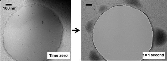
The molecular- and nano-scale structure of bulk materials can be imaged only by transmission electron microscopy (TEM), a technique that requires high vacuum (low pressure). Until now, this constraint made TEM incompatible with ‘wet’ materials, such as molecules dispersed in a liquid or gels swollen by a solvent. Working at the Materials Research Science and Engineering Center (MRSEC) on Polymers at UMass Amherst, Hoagland, McCarthy, and Russell successfully imaged the fine structure of wet polymeric materials prepared with ionic liquids. These non-volatile liquids dissolve or solvate an interesting spectrum of ‘soft’ polymer systems of fundamental and technological interest. A key objective is to refine the approach so that the dynamics of such systems can be imaged in situ and in real time, accomplishing tasks akin to those known from optical microscopy but at much smaller length scales. Shown below are two images, taken 1 second apart, of an ionic liquid spanning a 1-mm opening in a carbon film. Suspended in the liquid are polymer-coated nanoparticle tracers. In the interval between images, the thin film, originally spread over the opening, has destabilized by breaking into ~100-nm droplets that distribute around the rim. Nano- and micro-scale wetting transitions are key to numerous technologies ranging from microfluidics to ultralyophobic surfaces, but these transitions have never been visualized experimentally.